Definition: A minimally invasive procedure to visualize the coronary arteries using X-ray and contrast dye. Access is gained through the femoral artery in the groin.
Procedure Steps:
1. Preparation & Anesthesia: The groin area is cleaned and shaved. Local anesthesia is injected to numb the area.
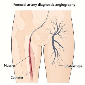
2. Arterial Access: A needle is used to puncture the femoral artery. A sheath (a short hollow tube) is inserted into the artery.
3. Catheter Insertion: A long, thin, flexible catheter is threaded through the sheath and up the aorta to the openings of the coronary arteries.
4. Injection of Contrast Dye: Contrast dye is injected through the catheter into the coronary arteries.
5. X-ray Imaging: X-ray movies (angiograms) are taken as the dye flows through the arteries, revealing any blockages or narrowings.
6. Removal: The catheter and sheath are removed. Pressure is applied to the site for 15-20 minutes to prevent bleeding.
Purpose / Benefits:
• Purpose: The “gold standard” for diagnosing Coronary Artery Disease (CAD).
• Benefits:
o Provides highly detailed images of blockages, their location, and severity.
o Can be used to guide further treatment like angioplasty or stenting.
Pre-procedure Tests (Creat, HBV/HCV):
• Creatinine (Creat): To check kidney function before using contrast dye, which can be harmful if kidneys are impaired.
• HBV/HCV: Screening for Hepatitis B and C for infection control and safety of healthcare workers and the patient.